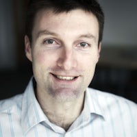
Dr James Bell
Research Associate
Overview
I am a researcher working within the Structural Biophysics Group and carry out a small amount of teaching on the undergraduate and postgraduate optometry courses.
Biography
Qualifications
2006: BSc Mathematics and Physics, Class I, University of Exeter
2010: PhD Physics, 'The relationship between the structure and mechanical properties of articular cartilage'.
Career overview
2019 - present: Research Associate, Cardiff University.
2018 - 2019: Teacher, Cardiff University.
2015 - 2018: Research Associate, Cardiff University.
2010 - 2015: Associate Research Fellow / Research Fellow, University of Exeter
Honorary appointments
2019 - present: Visiting Scientist, Diamond Light Source
2018: Visiting Lecturer, Universite Grenoble-Alpes, France.
2015 - present: Visiting Scientist, Exeter University
Honours and awards
2021: Accepted on to Cardiff Futures training scheme.
2020: Accepted on to Welsh Crucible future leaders training scheme.
2019: STEM certificate for 50 or more volunteering hours.
2016: Am J Physiol Heart Circ Physiol. work featured on journal cover.
2012: 62nd British Microcirculation Society Meeting - 2nd Joint Meeting with the American Microcirculation Society. 'Structural changes in loaded blood vessels'. Best Technology Abstract.
Speaking engagements
2020: S4SAS invited talk. "Nanomechanics of the ocular coat and the I22 biomechanics facility". Virtual conference.
2018: BioMedEng18 conference "A subfibrillar deformation mechanism in corneal collagen that affords flexibility". Imperial College London.
2017: "DIAMONDS" invited seminar. "Ocular structure and function in normal and pathological conditions". Diamond Light Source
2017: OBCS conference invited talk. 'The Hierarchical Response of Corneal Collagen to Load'. University of Southampton
2015: British Association for Surgery of the Knee Spring Conference. 'Morphological and chemical changes in human articular cartilage following RF coblation chondroplasty'. The International Centre, Telford
2014: British Microcirculation Society Young Investigators Conference. 'Nonlinear microscopy as a probe of blood vessel micromechanics'. University of Warwick.
Committees and reviewing
School committees
- Chair the Green Impact team
- Environment representative on the Health and Safety committee
- Member and "local champion" on IT committee
Reviewing
- Regular reviewer for Acta Biomaterialia
- Grant reviewer for
Publications
We are currently unable to retrieve the list of publications. Visit our institutional repository.Overview
My research involves quantifying the mechanical properties of tissue, and relating my findings to the micro- and nano-structure. Most tissues derive their mechanical properties from collagen, which forms an extracellular matrix tailored to the tissue function through interactions with proteoglycan, elastic fibres and interstitial fluid. An excellent paper on the role of collagen in the cornea can be found here. I use techniques such as multiphoton microscopy (part of the excellent bioimaging suite within VSBL) and X-ray scattering (predominantly at Diamond Light Source, the UK synchrotron) to obtain structural information about tissue that ranges in scale from molecular all the way up to whole eyes. I combine these techniques with mechanical tests using bespoke apparatus to visualise how the structures I see respond to stress.
Tropocollagen springs
My work in the field of ocular biomechanics led to a breakthrough in the understanding of collagen mechanics in general. In this paper I showed that some collagen fibrils are able to stretch significantly under relatively small stresses, due to a spring-like straightening of their supramolecular structure. This could have profound implications for our biomechanical understanding of not just the eye, but also blood vessels, skin, and many other tissues that exhibit this spring-like architecture (see an excellent paper by Ottani et al. for an overview).
Microvascular remodelling in diabetes
I worked on a British Heart Foundation sponsored project that investigated changes in our small arteries caused by diabetes. Our small arteries (approx 100 - 400 μm in diameter) are extremely important, because they are the primary means by which our bodies control organ perfusion. I carried out a multiphoton study in healthy arteries that illustrated the different stress transfer mechanisms present that control the response to pressure changes, as well as the highly heterogeneous distribution of stress through the vessel wall. I followed this up with an article investigating the changes associated with diabetes, which found arteries less able to distend and morphologically distorted due to the presence of pathologically thick taut bundles of collagen constraining the outer edge of the vessel.
Collaboration
I am actively seeking new collaborations with biologists who have an interest in tissue structure and/or biomechanics. If you think there may be overlap between our fields please drop me a line.